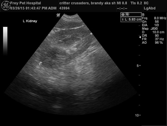
The use of ultrasound in small animal veterinary medicine has grown rapidly at Frey Pet Hospital. This is a non-invasive way to make an early diagnosis and allows for better management of diseases. Without the aid of ultrasound, these diseases previously went undetected for months to even years.
What is ultrasound?
An ultrasound examination is an imaging technique in which deep structures of the body can be visualized by recording echoes of ultrasonic waves directed into the tissues. Unlike x-rays which involve some small degree of radiation, ultrasound waves are considered to be entirely safe.
What is the difference between ultrasound and other types of imaging?
Because the ultrasound obtains a moving image, one can see precisely how the body is functioning in a rapid, non-invasive way. We can check the size, structure, and appearance of “the inside” of internal organs to see how each one is working in action.
Which parts of the body do you ultrasound?
The list of organs that can be evaluated with ultrasound is long! Most people are familiar with ultrasound for pregnancy diagnosis, however, we use ultrasound to detect detached retinas in the eye, check the thyroid, and visualize almost all other major organ systems. The basic abdominal ultrasound examines the liver, spleen, stomach, kidneys and urinary bladder, although most of the time, we are also evaluating the gall bladder, intestines, lymph nodes and prostate. Finding tumors and/or fluid in the abdomen and thorax is also an important part of the ultrasound exam.
What about the heart?
Using the ultrasound to evaluate a pet’s heart has a more specific name, called an echocardiogram. An echocardiogram offers tremendous detail about how the heart is functioning. It will reveal problems with heart valves, heart chamber dilation or thickening, as well as tumors located on or around the heart. The most important part of the echocardiogram is that it captures images of the heart in motion; we can use color flow doppler and pulse-wave doppler to study murmurs and other defects. This assessment allows for early medical (or even surgical) intervention, prolonging a pet’s quality and quantity of life.
Does the technique have any drawbacks?
Ultrasound examinations are of little value in the examination of organs that contain air. Ultrasound waves will not pass through air and therefore it cannot be used to examine the lungs.
Do you need to be specially trained to use ultrasound?
Yes, the machine is totally operator dependent. Dr. Jennifer Feuerbach has spent several years developing her skills, and enjoys attending continuing education in ultrasound whenever possible. We have invested in the most advanced ultrasound technology available in the veterinary market today. We also have the capability of consulting with board-certified cardiologists and radiologists quickly in the event of an unusual ultrasound case.


